‏إظهار الرسائل ذات التسميات سنة اولى تكليف. إظهار كافة الرسائل

٢٠‏/٠٦‏/٢٠٠٨

atropine and atropine overdose


i writ about atropin and atropine over dose because ihad astory with acase of atropine over

sign and symptoms


include vasodilation;

drying of the mouth;

increase in pulse rate;

inhibition of contractions of the gastrointestinal tract, ureter, and bladder;

and reduction of salivary, bronchial, gastric, and sweat gland secretions.

Following clinical and larger doses, atropine sulfate causes dilation of the pupils (mydriasis) and paralysis of accommodation (cycloplegia) and, in narrow-angle

glaucoma, can increase intraocular pressure


0.5 mg -

Slight dryness of nose and mouth; bradycardia
1 mg

- Greater dryness of nose and mouth with thirst; slowing, then acceleration of heart; slight mydriasis.
2 mg -

Very dry mouth; tachycardia with palpitations; mydriasis, slight blurring of near vision; flushed, dry skin.
5 mg

- Increase in above symptoms plus disturbance of speech; difficulty in swallowing; headache; hot, dry skin; restlessness with asthenia.
10 mg and over

- Above symptoms to extreme degree plus ataxia, excitement, disorientation, hallucinations, delirium, and coma.


Contraindications
Glaucoma; adhesions (synechiae) between the iris and lens of the eye;

asthma;

Sal-Tropine is contraindicated in patients with an allergy to atropine or to sulfate.


treatment of Atropine Overdosage

a short acting barbiturate or diazepam may be given as needed to control marked excitement and convulsions.


Large doses for sedation should be avoided because central depressant action may coincide with the depression occurring late in atropine poisoning. Central stimulants are not recommended.


Physostigmine, given as an atropine antidote by slow intravenous injection of 1 to 4 mg (0.5 to 1.0 mg in children), rapidly abolishes delirium and coma caused by large doses of atropine. Since physostigmine is rapidly destroyed, the patient may again lapse into coma after one to two hours, and repeated doses may be required.


Artificial respiration with oxygen may be necessary.


Ice bags and alcohol sponges help to reduce fever, especially in children


.


١٨‏/٠٦‏/٢٠٠٨

unilatteral hand swelling

Today I meet in the primary health care that I work in it a strange case for me
I mean that I didn’t such this case before
If any one have any idea about the differential diagnosis of this case I will be too happy if he help me

This is the brief history

Middle aged female patient come to primary health care unit complaining of unilateral hand swelling from 2dayes gradual in course
There no history previous trauma , no history chronic illness , no history of previous operation


???????????????????????

٠٢‏/٠٥‏/٢٠٠٨

قصة ال ICDL

كلنا طبعا عارفين اهمية شهادة ال
ICDL بذات الايام اللى احنا عايشن فيها
مفيش حد مثلا يقدر يحضر ماجستير او دكتوراة من غيرها غير اهميتها لخرجين تجارة وهندسة و .....و......
وعشان كل هذة الاسباب

انا قررت انى احصل على هذة الشهادة
اولا حاجة عملتها انى سالت واستفسرت عن الكتب والامتحانات وطريقة الامتحان وسكور النجا ح وبعد ان جمعت البيانات

بدات فعلا فى التنفيذ والحمد لله
اول حاجة الكتب باللغة الانجليزية V4 التحميل من الموقع دا
الشرح
بس كان فى عندى مشكلة انى مفيش عندى صير على القراة فققررت ان ابحث عن مصدر للشرح لتسهيل على نفسى
الحمد لله ربنا وفقنى واستطعت ان اصل الى هذا الموقع الرائع
اما الامتحانات
النجاح من 75%
والموقع دا ممكن منه تحميل الامتحانات

على فكرة الامتحانات الموجودة فية قريبة جدا من الامتحان الحقيقى
وربنا يوفقك الجميع

٢٤‏/٠٤‏/٢٠٠٨

SCABIES

DIAGNOSIS
The combination of a history of pruritus (especially at night), a classic rash, and itching in household or sexual contacts is adequate for the diagnosis of scabies. Mites seen on microscopic examination of skin scrapings confirm the diagnosis


TREATMENT OF CLASSIC SCABIES
Topical Agents. In adults and children over five years of age, 5 percent permethrin cream (Elimite) is standard therapy for scabies


When a scabicide is prescribed, the patient (or a parent or other caregiver) should be informed that itching may persist for up to four weeks after successful treatment. It may take that long for the skin to slough residual mite debris and for the allergic reaction to subside. Itching may be managed with antihistamines and, if necessary, the addition of a topical steroid. However, steroids should not be prescribed before the completion of primary therapy

Because some developing louse larvae may survive initial treatment, a second course of

treatment is recommended seven to 10 days latter


. .
Environmental Measures.

Once scabies has been diagnosed, the physician should explain its basic epidemiology. Otherwise, medication alone might be relied on to eradicate the infestation. If environmental control measures are not instituted, treatment failure will occur, because mites are able to survive and reinfest the patient. Thus, it is critical to decontaminate all linens, towels, and clothing used in the previous four days by hot-water washing (60°C) and heated drying. Items that cannot be washed in hot water should be dry cleaned or sealed in a plastic bag for five days. Even if household and sexual contacts have no symptoms, they should follow the same cleaning

procedures. Treatment must be simultaneous for al


to more information visit

١٣‏/٠٤‏/٢٠٠٨

Amebiasis ,treatment of Nematode Infections

Albendazole (Albenza-
Used for hookworm, pinworm, and roundworm. Decreases ATP production in worm, causing
energy depletion, immobilization, and, finally, death
Adult Dose
Ascariasis, enterobiasis, hookworm, or trichuriasis: 400 mg PO as single dose, may repeat in 3 wk if necessary
Strongyloidiasis: 400 mg PO qd for 3 d, may repeat regimen in 3 wk if necessary
Trichinosis: 400 mg PO bid for 15 d
Pediatric Dose
Ascariasis, enterobiasis, hookworm, or trichuriasis<2> 200 mg PO as single dose, may repeat in 3 wk if necessary>2 years: Administer as in adults
Strongyloidiasis<2> 200 mg PO qd for 3 d, may repeat regimen in 3 wk if necessary>2
years: Administer as in adults
/R
alzental tablet 200mg
single dose repeated in 3 weeks
childern more than 2y
\R
alzental susp 200mg
2bottle as single dose repeated in 3 weeks
for more details visit the link

Amebiasis
Metronidazole (Flagyl)
Description
Active against various anaerobic bacteria and protozoa. Appears to be absorbed into cells. Intermediate metabolized compounds are formed and bind DNA and inhibit protein synthesis, causing cell death. Antimicrobial effect may be due to production of free radicals.Indicated for invasive E histolytic infections.
Adult Dose
500-750 mg PO tid for 10 d
Pediatric Dose
35-50 mg/kg PO divided tid for 10 d
Contraindications
Documented hypersensitivity
Interactions
Cimetidine may increase toxicity of metronidazole; may increase effects of anticoagulants; may increase toxicity of lithium and phenytoin; ingestion of ethanol during therapy may induce a disulfiramlike reaction with abdominal cramps, nausea, and emesis
Pregnancy
B - Usually safe but benefits must outweigh the risks.
Adjust dose in hepatic disease; monitor for seizures and development of peripheral neuropathy; alcoholic beverages should be avoided during
Precautions
administration and for 3 d after

٢٨‏/٠٧‏/٢٠٠٧

neonatal

Majority of the newborns do not require any form of intervention at birth. However, approximately 10% need some assistance to begin breathing and 1% requires extensive resuscitation


Initial Assessment

At birth the following characteristics are rapidly assessed to identify the need for resuscitation:

(a) Is the baby born after full term gestation?

(b) Is the amniotic fluid clear of meconium and evidence of infection?

(c) Is the baby breathing or crying?

(d) Does the baby have good muscle tone?


If the answer to all the questions is positive, the newborn does not require resuscitation.


  • To prevent heat loss, the baby should be dried and covered in linen to maintain body warmth.

  • In preterm babies, especially those with gestation age <28>

  • The newborns' color, breathing and activity should be continuously assessed.


If the answer to any one of the above questions is negative, the neonate would require one or more of the following actions in sequence:


A. Initial steps in stabilization (clearing the airway, positioning, stimulating)


B. Ventilation


C. Chest compressions


D. Medication/s and volume expansion.


Progress to the next step in the sequence is based on simultaneous assessment of the three vital signs:


  • respiration,

  • heart rate

  • and color.

Approximately 30 sec have been allocated to complete one step successfully,


re-evaluate and decide whether to progress to the next step.


Respiratory activity The neonates' breathing should be checked.


  • Rate,
  • depth
  • and symmetry of respiration should be evaluated.
  • Any abnormal breathing patterns such as gasping or grunting should be noted.

  • Heart rate Palpation of the pulse below the umbilical cord has been found to be reliable only if heart rate is more than 100 beats/minute. [4]
  • Heart rate is best evaluated by auscultation.

  • Colour Although a healthy baby is born blue, the color becomes pink within 30 seconds of the start of effective breathing.
  • A healthy neonate achieves and maintains pink mucous membranes without supplementary oxygen.
  • Evaluation of central cyanosis is done by examining the face, trunk and mucous membranes. Peripheral cyanosis is often observed and does not by itself indicate hypoxemia.
  • Pallor or mottling is seen in neonates having decreased cardiac output, hypovolemia, severe anemia, hypothermia or acidosis.

  • Tone If the neonate is very floppy, it is most likely unconscious and would require respiratory support.

to read more go to

http://www.ijccm.org/article.asp?issn=0972-5229;year=2007;volume=11;issue=2;spage=81;epage=89;aulast=Kapoor


٠٩‏/٠٧‏/٢٠٠٧

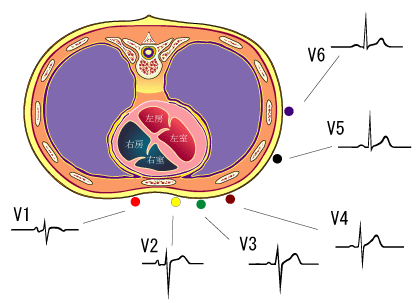
ECG CHANGE WITH ISHEMIC HEART


V1,V2 represent right ventricle


V3,V4 represent septum of heart


V5,V6 represent left ventricle


Site of infarctionThe ECG has been used to localize the site of ischemia and infarction. Some leads depict certain areas; the location of the infarct can be detected fairly accurately from analysis of the 12-lead ECG. Leads that best detect changes in commonly described locations are classified as follows:


  • lead II,III, aVF.......................> represent inferior wall MI

  • lead V1 and V2 .......................> septal

  • lead I, aVFL.............................>represent latteral wall MI

  • lead V1,V2................................>represent strict anterior

  • lead V1,V2,V3,V4....................> anterio septal

  • lead V1 TO V6.........................> extensive anterior

The classic changes of necrosis (Q waves), injury (ST elevation), and ischemia (T wave inversion) may all be seen during acute infarction.


In recovery,


  • the ST segment is the earliest change that normalizes,

  • then the T wave;

  • the Q wave usually persists.

Therefore, the age of the infarction can be roughly estimated from the appearance of the ST segment and T wave.


The presence of the Q wave in the absence of ST and T wave abnormality generally indicates prior or healed infarction


ECG

P wave

The P wave represents the wave of atrail depolarization

p-R interval

The period of time from the onset of the P wave to the beginning of the QRS complex
normally ranges from 0.12 to 0.20 seconds in duration or(3-5 smal sequre)

represents the time between the onset of atrial depolarization and the onset of ventricular depolarization

Q wave
pathological Q wave means Qwave>1/3 R wave or >1 big sequre which ocurrce with old MI

QRS complex

The QRS complex represents ventricular depolarization.
The duration of the QRS complex is normally 0.06 to 0.1 seconds. (<2.5>abnormal duration
If the QRS complex is prolonged (> 0.1 sec), conduction is impaired within the ventricles.
This can occur with:

  • bundle branch blocks

  • a ventricular foci (abnormal pacemaker site) becomes the pacemaker driving the ventricle. Such an ectopic foci nearly always results in impulses being conducted over slower pathways within the heart, thereby increasing the time for depolarization and the duration of the QRS complex.
abnormal shape

the shape changes depending on

  • which recording electrodes are being used.

  • The shape will also change when there is abnormal conduction of electrical impulses within the ventricles


ST segment
The isoelectric period (ST segment) following the QRS is the time at which the entire ventricle is depolarized
The ST segment is important in the diagnosis of ventricular ischemia or hypoxia because under those conditions, the ST segment can become either depressed or elevated.

T wave
The T wave represents ventricular repolarization
Sometimes a small positive U wave may be seen following the T wave
This wave represents the last remnants of ventricular repolarization.
Inverted or prominent U waves indicates underlying pathology or conditions affecting repolarization.

Q-T interval
The Q-T interval represents the time for both ventricular depolarization and repolarization to occur, and therefore roughly estimates the duration of an average ventricular action potential. This interval can range from 0.2 to 0.4 seconds depending upon heart rate.
At high heart rates, ventricular action potentials shorten in duration, which decreases the Q-T interval.

Because prolonged Q-T intervals can be diagnostic for susceptibility to certain types of tachyarrhythmias, it is important to determine if a given Q-T interval is excessively long.
In practice, the Q-T interval is expressed as a "corrected Q-T (QTc)" by taking the Q-T interval and dividing it by the square root of the R-R interval (interval between ventricular depolarizations). This allows an assessment of the Q-T interval that is independent of heart rate.
Normal corrected Q-Tc intervals are less than 0.44 seconds.
  • cause LQTS include the following:
  • Drugs (many antiarrhythmics, tricyclics, phenothiazines, and others)
  • Electrolyte abnormalities ( K+, Ca++, Mg++)
  • CNS disease (especially subarrachnoid hemorrhage, stroke, trauma)
  • Hereditary LQTS (e.g., Romano-Ward Syndrome)
  • Coronary Heart Disease (some post-MI patients)
  • paient may be develop arrythmia (dr m)


٠٢‏/٠٧‏/٢٠٠٧

How to do CPR and use an AED

its too important to any medical persone to learn to do CPR

Insulin injections

Diabetes is common health problem


Guide to Insulin Injections

    1. In general, insulin shots can be given pretty much wherever there’s enough fat under the skin. The main areas are the abdomen, thighs, and the back of the upper arms. The abdomen is generally used the most. It’s easy to reach, and the insulin is absorbed from the abdomen at a more consistent rate from shot to shot.
    1. Each injection site should be about the size of a quarter. So you only have to move about a finger-width away from your last shot each time you rotate
    1. Don’t inject too close to moles or scars.

    2. If you inject in the arm, use only the outer back area of the upper arm (where the most fat is). Otherwise, you run the risk of injecting into a muscle.
    3. If you inject in the thigh, use only the top and the outside area. Stay away from the inner thighs—rubbing between the legs can make the injection site sore.
      If you inject in the abdomen, don’t do it too near the navel. The tissue there is tougher and makes the insulin absorption less predictable.
    4. Push the plunger more slowly while injecting the insulin.
      Count to 5 or 10 after pushing in the plunger and before removing the needle.
    5. Check the angle of the needle. You may need to straighten it a little (to a 90° angle).
    6. Check the injection site for lumpiness. If it’s lumpy, choose another site.

Insulin Storage

  • Good insulin care begins with how the insulin is stored. Insulin does not work well when it’s kept for too long or is exposed to extreme temperatures
  • stay fresh for up to month without refrigeration

  • If you would rather keep all insulin in the refrigerator, make sure to warm up the insulin before injecting it. Cold insulin can make the shot uncomfortable. Draw up the right amount into the syringe, then roll the syringe gently between your hands until it feels warm.
  • Always check the insulin before you use it. Rapid- and short-acting insulin should look clear. There should be no cloudiness, little bits floating in the liquid, or change in color. Intermediate- and long-acting insulin should look cloudy, but you should not see any large clumps floating around. If you see any of these signs, discard the bottle.


  • Push the plunger more slowly while injecting the insulin.
    Count to 5 or 10 after pushing in the plunger and before removing the needle.
  • Check the angle of the needle. You may need to straighten it a little (to a 90° angle).
  • Check the injection site for lumpiness. If it’s lumpy, choose another site.


">for more reading