


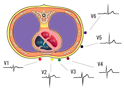
V1,V2 represent right ventricle
V3,V4 represent septum of heart
V5,V6 represent left ventricle
Site of infarctionThe ECG has been used to localize the site of ischemia and infarction. Some leads depict certain areas; the location of the infarct can be detected fairly accurately from analysis of the 12-lead ECG. Leads that best detect changes in commonly described locations are classified as follows:
- lead II,III, aVF.......................> represent inferior wall MI
- lead V1 and V2 .......................> septal
- lead I, aVFL.............................>represent latteral wall MI
- lead V1,V2................................>represent strict anterior
- lead V1,V2,V3,V4....................> anterio septal
- lead V1 TO V6.........................> extensive anterior
The classic changes of necrosis (Q waves), injury (ST elevation), and ischemia (T wave inversion) may all be seen during acute infarction.
In recovery,
- the ST segment is the earliest change that normalizes,
- then the T wave;
- the Q wave usually persists.
Therefore, the age of the infarction can be roughly estimated from the appearance of the ST segment and T wave.
The presence of the Q wave in the absence of ST and T wave abnormality generally indicates prior or healed infarction


ليست هناك تعليقات:
إرسال تعليق